Our mission is to cure complex brain diseases. We are reinventing neuro drug discovery by combining human brain models, scaled biology and machine learning to systematically discover and develop novel drugs with greater therapeutic potential.
Cures, not correlations.
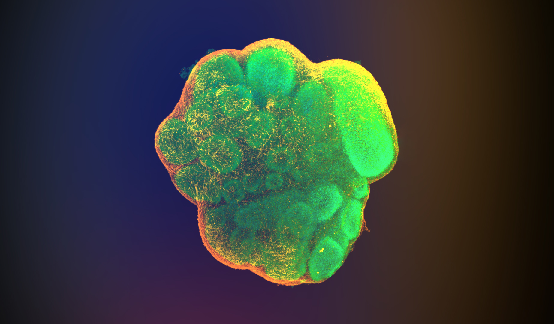
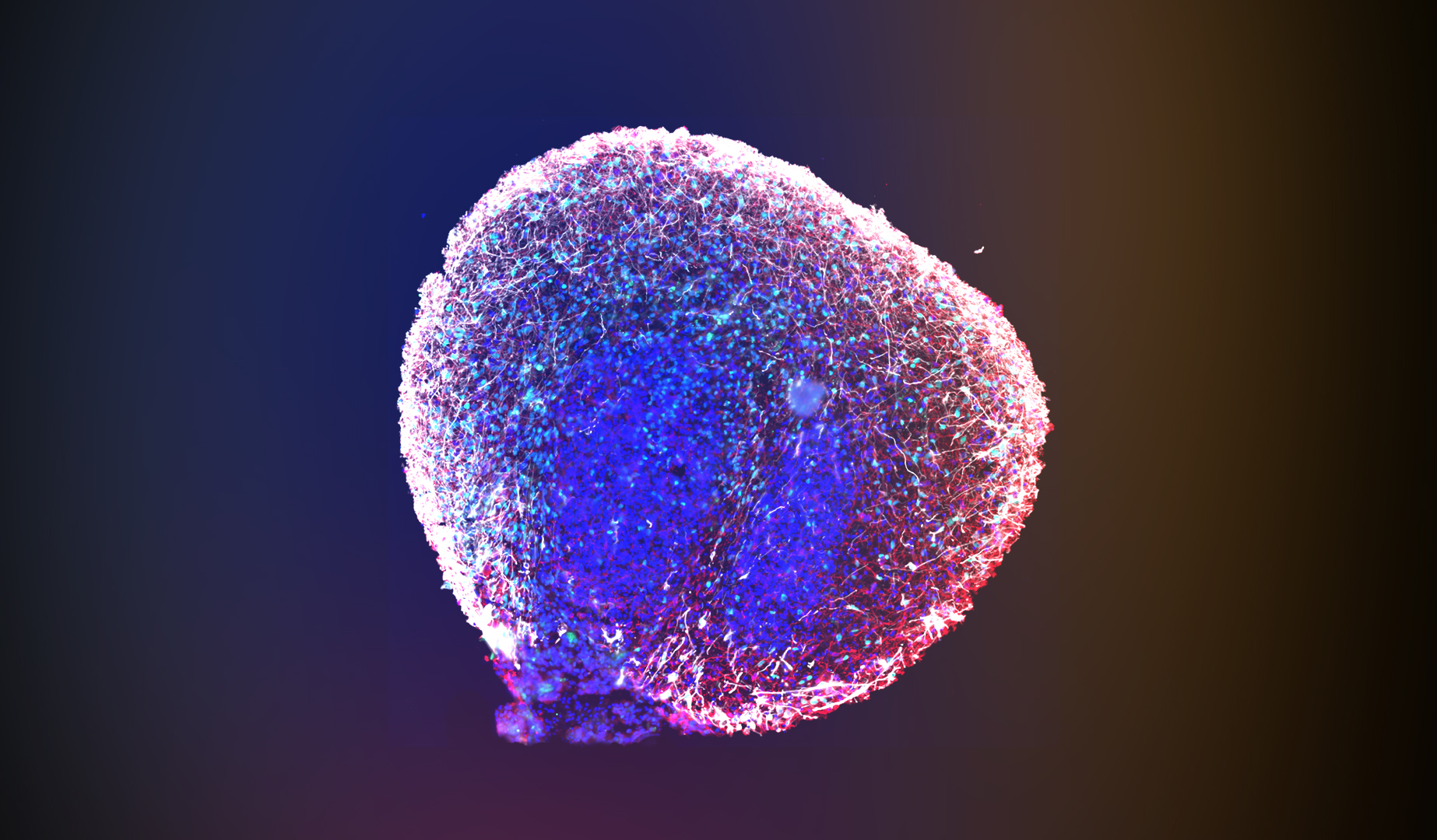
Confocal 3D image of a clarified, immunofluorescence labeled cerebral organoid at Day 35 showing MAP2 (orange/yellow) and SOX2 (green). Day-35 cerebral organoids show emerging outer neuronal zones and large internal stem cell zones generating neurons and glial cells. The stem cell zones are the internal globe-like collections of brightly labeled cells (SOX2). Newborn neurons (MAP2+) are the stringy cells on the surface.
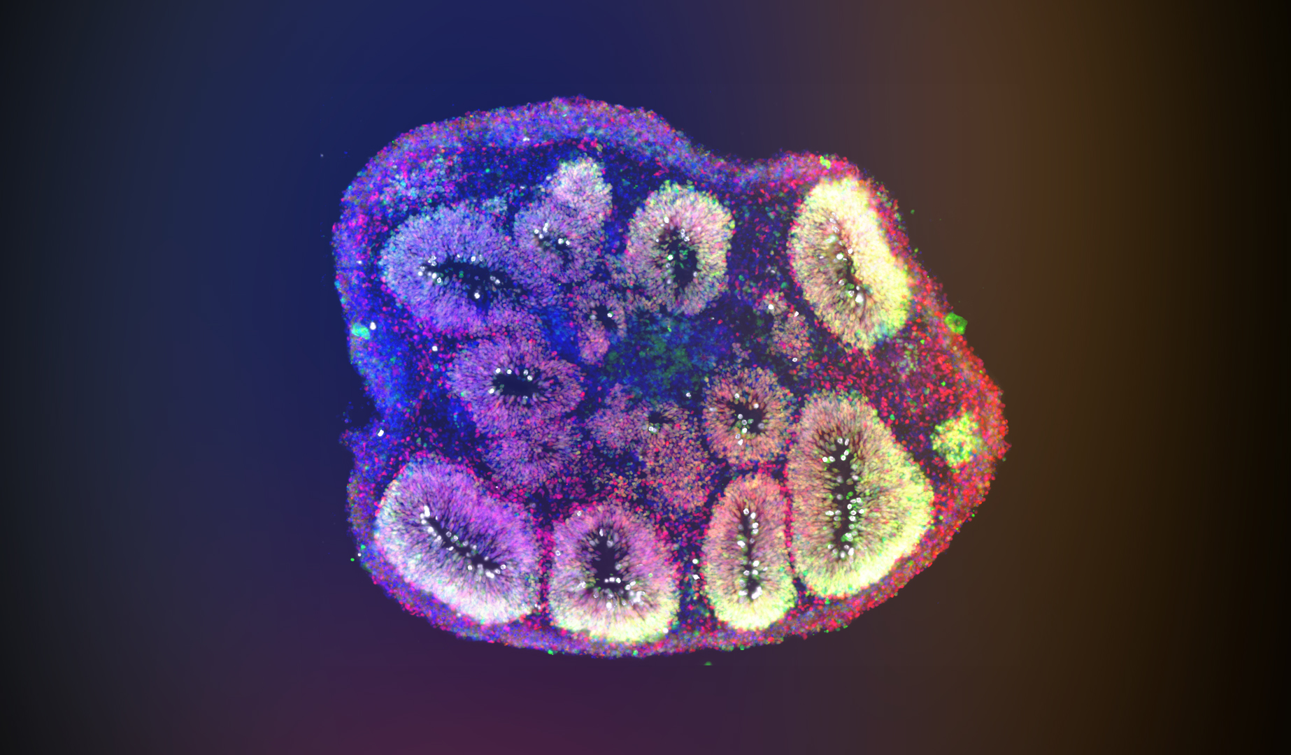
Immunofluorescent labeling for Emx1 (red) and Sox2 (green), with DAPI, in a tissue cross section at Day 35. Emx1+ cortical neurons migrate away from the buds and accumulate near the organoid surface, forming a cell-dense region similar to the cortical plate of developing mammalian brains.
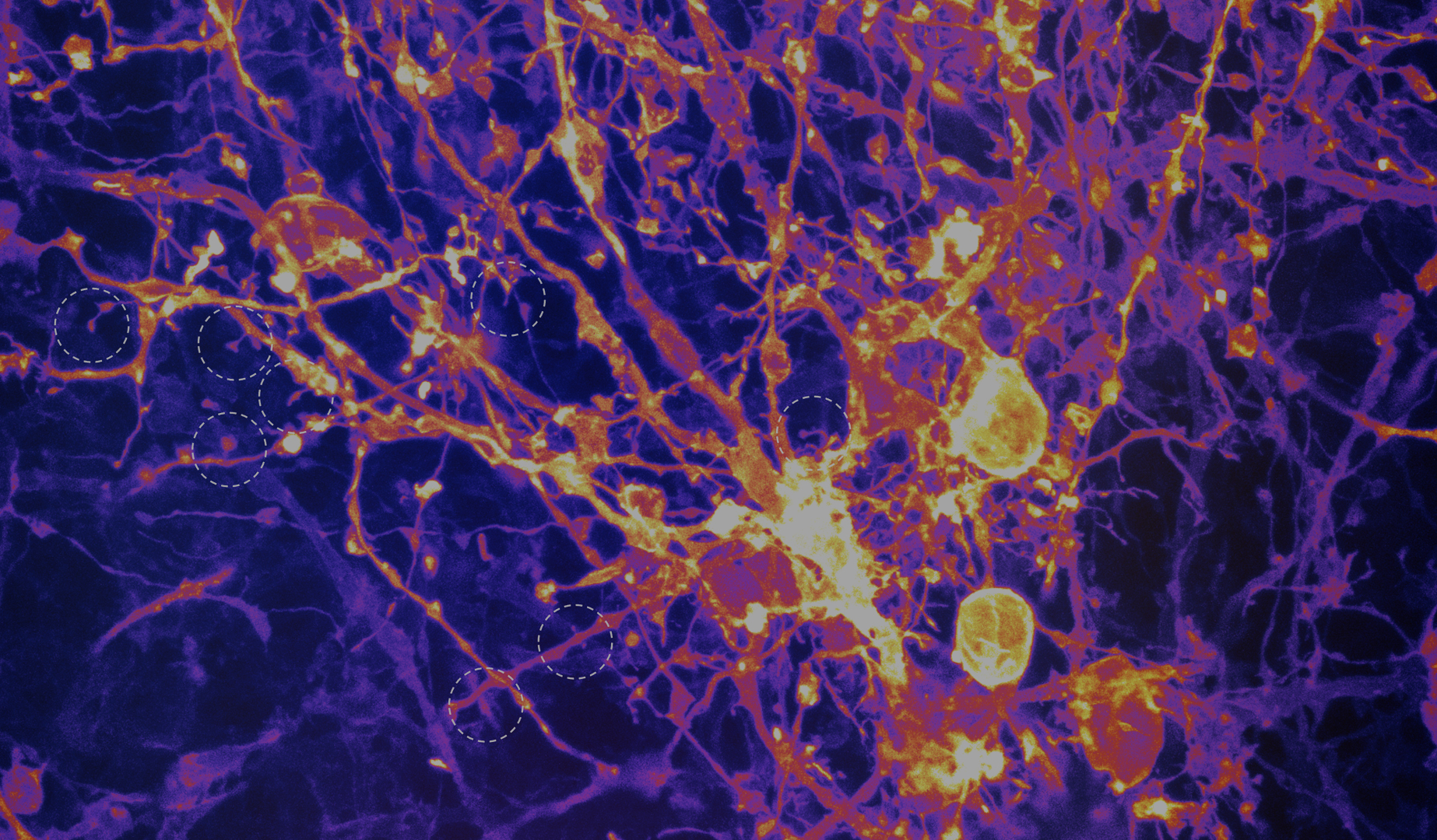
High-resolution confocal 3D image of neurons labeled with DiI (a lipophilic dye that labels cell membranes). Tiny synaptic structures called dendritic spines are formed by neurons that support synaptic transmission. Spines (dotted circles) contain neurotransmitter receptors that are the information receivers of the post-synaptic cell.
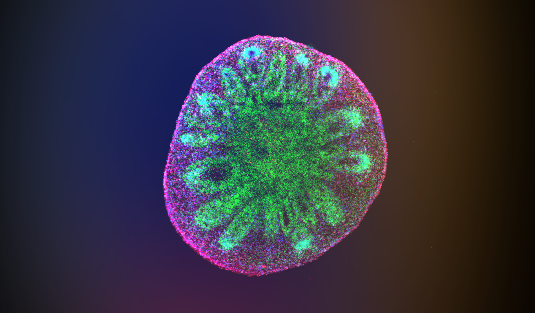
Immunofluorescent composite image of a tissue cross section, showing MAP2, NeuN, and DAPI. Day 90 organoids show well defined outer neuronal zones and remnant internal stem cell zones generating neurons and glial cells. The neuronal zones are functionally and anatomically interconnected by neurites (MAP2+ dendrites harboring active synapses).
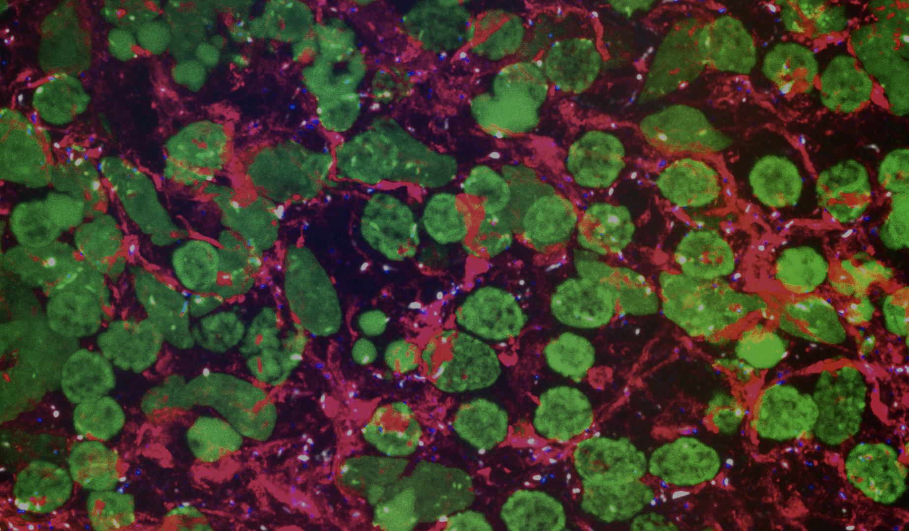
Forebrain neurons show extensive MAP2+ neurites (red), dotted with tiny white puncta (synapsin 1) and blue puncta (gephyrin). Synapsin 1 and gephyrin often form “doublets”, marking the pre-synaptic and post-synaptic portions of each synapse. The large green globes are individual cellular nuclei.
(the thin neuronal and glial processes and synapses filling the space between neuronal cell bodies). Most of the cortical gray matter volume in mammals is occupied by neuropil —increasingly so in primates— which is reproduced by organoids. This system allows the detailed investigation of 3D neuropil metrics, including neurite/cellular density according to cell type, as well as synapse density measurements, in healthy and diseased patient-derived organoids.